

Discover PhysioFlow
The Time Bomb !

The Time Bomb !
What do we measure?
Cardiac Output (CO) is the amount of blood pumped by the heart in liters per min. It is the true "raison d'être" of the heart.
CO is a hemodynamic parameter that plays a key role in several physiological equilibriums. For instance: it is the dynamic force that generates arterial blood pressure by combination with the systemic vascular resistance (SVR). Another example is the close interaction between Co, oxygen consumption (VO2) and peripheral oxygen extraction described by the "Fick" equation.
As a result and for instance, systemic hypertension should not be managed without the knowledge of cardiac output because CO is a key contributor to the calculation of SVR which is essential in understanding and managing this pathology. Similarly, relying on VO2 only to evaluate the physiology of exercise is way too restrictive considering the importance of cardiac and peripheral parameters as potential limiting factors for human performance.


CO, Stroke Volume (SV) and their components (preload, contractility, afterload) need to be evaluated in dynamic manner. For instance, the rate of increase of SV and contractility during exercise differs considerably from a heart failure patient to an athlete. Abnormal trend patterns in SV during exercise are proven signs of cardiac function impairment (e.g. coronary artery disease). Another example: In critical care, fluid management is of paramount importance. Evolution and variation of SV are markers of fluid status and need, for instance during Passive Leg Raise test.
Very unfortunately CO has so far been underused because of the invasive, cumbersome and inaccurate nature of the available measurement techniques. Their cost has also been a limiting factor.
How do we measure it?
PhysioFlow® redefines hemodynamic monitoring
Accurate, noninvasive hemodynamics have been the Holy Grail sought after for four decades. When conventional impedance cardiology appeared, many felt the goal was reached, only to be disappointed by the limitations that quickly became apparent:
.Poor correlation to invasively-determined values
.Patient-related limitations (obesity, emphysema, pulmonary edema, mechanical ventilation etc.)
.Inaccuracies related to motion artifact and electrode position
.Costs sometimes prohibitive
Over the last two decades, conventional (Z0-based) ICG has been improved and tweeked. However its fundamental dependence on the unreliable baseline thoracic impedance (Z0) could never be overcomed. And Z0 proved to be random value in too many cases. To solve the problem, impedance cardiology was reinvented by PhysioFlow to be independent of baseline thoracic impedance (Z0).
The breakthrough that accomplished this was to use the morphology of the impedance signal (pure waveform analysis), instead of its baseline value, a patented-approach called Signal-Morphology Impedance Cardiology (SM-ICG™).
SM-ICG™ provides accurate, reproducible and sensitive measurements even in challenging measurement conditions and difficult patients, pushing the limits of noninvasive hemodynamic monitoring.
.What is impedance cardiography ?
Impedance cardiography (ICG), also referred to as thoracic electrical Bioimpedance (TEB) and electrical impedance plethysmography (EIP), has been researched since the 1940's. With ICG, disposable sensors on the neck and chest are used to transmit and detect electrical and impedance changes in the thorax, which are used to measure and calculate hemodynamic parameters.
.How does ICG Work ?
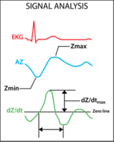
A low magnitude high frequency alternative current is transmitted through the chest. ICG™ measures the Baseline Impedance (Resistance or Z0) to this current together with variations induced by the respiration and the cardiac flow.
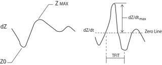
The sensed impedance signal (delta Z or dZ) is then processed and analyzed together with its derivative over time (dZ/dt) a mirror image of dP/dt. Both signals are pulsatile waveforms.
.How does standard ICG (BioZ, Medis, Osypka and Nicas etc ...) compute Stroke Volume ?
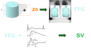
Standard ICG filters out the respiratory component of the impedance signal but uses the impedance baseline in the equation to compute Stroke Volume (SV) and subsequently Cardiac Output (CO = SV X Heart Rate).
The impedance baseline (Z0) is combined with an estimation of the shape/volume of the thorax, and L (length) or distance between electrodes to estimate thoracic fluid content (TFC). The TFC is used as a reference to analyze the pulsatile components of the signals (dZ and dZ/dt).
The pulsatile waveform is indeed the part of the impedance signal that is in motion in accordance with the blood flow generated by the heart.
.What are the limitations of standard ICG ?
Standard ICG works only in normal or near normal patients and exclusively at rest or very moderate exercise. Obesity, pulmonary edema, emphysema, ventilation are some known limitations to ICG. It is very sensitive to electrodes placement and therefore not very reproducible from one operator to another.
.What is the origin of these limitations ?
There are two main causes for these limitations
The impedance signal is of very low magnitude (1/1000 of the magnitude of an EKG signal) and therefore its sensitivity to interferences and artifacts is high.
The Z0 parameter, which is key in the classic equations, is highly inaccurate when the patient is not normal or near normal, not at rest or when electrode placement is sub-optimal.
.How to improve the robustness/stability of the signal ?
It is now possible to improve the stability and signal/noise ratio of impedance cardiography thanks to newly developed filtering techniques. Two methods have been offered on the market recently: SM-ICG™ (PhysioFlow®) and Bioreactance®. They have proven to advance the landscape in impedance cardiography.
.How does Bioreactance® work ?
The Bioreactance® approach is based on the assumption that when blood flows out of the heart, phase shifts are created in alternating radiofrequency electrical currents applied across the patients chest wall. Such phase shifts are conceptually similar to a Frequency Modulation, of FM, as used in FM radio transmissions. The phase shifts are measured continuously and have been shown to relate almost linearly to blood flow in the aorta. Just like FM and AM radios, Bioreactance® plays the same "impedance" music, but with a clearer sound.
.How do SM-ICG™ filters work ?
SM-ICG™ uses a first level optimized input filter finely adapted to the high frequency current transmitted,
so that the Impedance signal measured is mostly free from noise coming from other electronic or physiologic emitters.
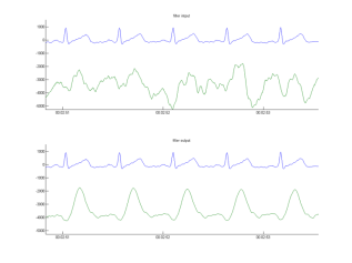
SM-ICG™ also uses a second level highly sophisticated filter, called HD-Z™ (for High Definition Impedance) to eliminate all artifacts from the chest impedance signals that are not correlated with the heart cycle and the generation of cardiac flow. With HD-Z™ the relative amplitude of the noise to the amplitude of the cardiac signal does not matter. It has been developed to cancel severe artifacts in the most demanding situations like a running race (marathon) or in a very noisy critical care environment. Below are two examples of how the filter works on "difficult" dZ signals
Example 1:
HD-Z™ applied to the dZ signal of a marathon runner (the green signal above is before filtering and the green
signal below is after HD-Z™ filtering)
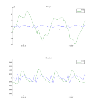
In these two cases the signal is totally stable and reproducible after application of the HD-Z filter.
.What are the similarities between Bioreactance® and SM-ICG™ ?
SM-ICG™ has been pioneering a radically new approach in ICG.
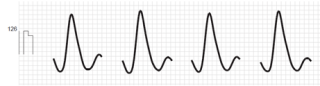
The calculation of SV is entirely based on a morphological/Differential analysis of the impedance waveform, factoring out the problematic impedance baseline (Z0) from the equation. The dZ and dZ/dt signals are electrical representations of the blood flow generated by the heart, just like a Doppler waveform is an ultrasound picture of the flow. It is immediately evident that a heart failure patient and an athlete present very different waveforms and that these waveforms contain the necessary information to compute SV. The designers of Bioreactance® have used the same approach ten years later and applied it to their "FM" signal, which looks identical to the SM-ICG™ signals when measurement conditions are optimal. Below is an example of Bioreactance® signals as an
illustration of that fact. Please compare this signal to the SM-ICG™ signals from above,
especially those of the ventilated ICU patient.
Example 2:
HD-Z™ applied to the raw dZ signal of a ventilated ICU patient
.What is the difference between Bioreactance® and SM-ICG™ ?
Just like FM and HD radios are two generations of radios, Bioreactance® and SM-ICG™ (HD-Z™) are two generations of impedance cardiograph signals filters. FM radio and Bioreactance® share the same set of flaws. In relation to its poor reception, FM radio contains cracks and distortion. Similarly, Bioreactance® is sensitive to a sub-optimal measurement environment. As a result, the Bioreactance® designers have limited it to a long one minute averaging time to guarantee there is an adequate amount of "good" beats to provide a visually stable average.
On the contrary, the HD-Z™ filter extracts the useful and clear information from a noisy signal in a dynamic manner, enabling shorter averaging times and more "information-rich" trends. PhysioFlow® has been proven to cancel noise from even extreme measurement environments like marathon races or maximal exercise testing of highly trained athletes. No other device has been successfully used in such difficult conditions and provides clinically useful information in such a dynamic fashion.
Another unique and published benefit of SM-ICG™ compared to other systems is that, thanks to the clarity, level of details and stability of its filtered signals, the practitioner can use it to see abnormalities on the waveforms that are signs of probable cardiac pathologies like ventricular dissynchrony, fluid overload, and ventricular stiffness. Therefore SM-ICG™ enables early detection of possible heart conditions that are potentially very detrimental to the patient if not treated on time.
.Conclusion
Bioreactance® and SM-ICG™ are two new generation ICG systems, applying similar concepts to improve the technology. SM-ICG™ has pioneered the pure morphological analysis of the signal, followed by Bioreactance®. SM-ICG™ applies a more advanced filtering technology with proven results in the most difficult measurement environments.
Last update : November 2022 - Contact - Legal - Manatec Biomedical
How do we measure it?
CO, Stroke Volume (SV) and their components (preload, contractility, afterload) need to be evaluated in dynamic manner. For instance, the rate of increase of SV and contractility during exercise differs considerably from a heart failure patient to an athlete. Abnormal trend patterns in SV during exercise are proven signs of cardiac function impairment (e.g. coronary artery disease). Another example: In critical care, fluid management is of paramount importance. Evolution and variation of SV are markers of fluid status and need, for instance during Passive Leg Raise test.
Very unfortunately CO has so far been underused because of the invasive, cumbersome and inaccurate nature of the available measurement techniques. Their cost has also been a limiting factor.
Cardiac Output (CO) is the amount of blood pumped by the heart in liters per min. It is the true "raison d'être" of the heart.
CO is a hemodynamic parameter that plays a key role in several physiological equilibriums. For instance: it is the dynamic force that generates arterial blood pressure by combination with the systemic vascular resistance (SVR). Another example is the close interaction between Co, oxygen consumption (VO2) and peripheral oxygen extraction described by the "Fick" equation.
As a result and for instance, systemic hypertension should not be managed without the knowledge of cardiac output because CO is a key contributor to the calculation of SVR which is essential in understanding and managing this pathology. Similarly, relying on VO2 only to evaluate the physiology of exercise is way too restrictive considering the importance of cardiac and peripheral parameters as potential limiting factors for human performance.
In these two cases the signal is totally stable and reproducible after application of the HD-Z filter.
.What are the similarities between Bioreactance® and SM-ICG™ ?
SM-ICG™ has been pioneering a radically new approach in ICG.
The calculation of SV is entirely based on a morphological/Differential analysis of the impedance waveform, factoring out the problematic impedance baseline (Z0) from the equation. The dZ and dZ/dt signals are electrical representations of the blood flow generated by the heart, just like a Doppler waveform is an ultrasound picture of the flow. It is immediately evident that a heart failure patient and an athlete present very different waveforms and that these waveforms contain the necessary information to compute SV. The designers of Bioreactance® have used the same approach ten years later and applied it to their "FM" signal, which looks identical to the SM-ICG™ signals when measurement conditions are optimal. Below is an example of Bioreactance® signals as an
illustration of that fact. Please compare this signal to the SM-ICG™ signals from above,
especially those of the ventilated ICU patient.
Example 2:
HD-Z™ applied to the raw dZ signal of a ventilated ICU patient
.What is the difference between Bioreactance® and SM-ICG™ ?
Just like FM and HD radios are two generations of radios, Bioreactance® and SM-ICG™ (HD-Z™) are two generations of impedance cardiograph signals filters. FM radio and Bioreactance® share the same set of flaws. In relation to its poor reception, FM radio contains cracks and distortion. Similarly, Bioreactance® is sensitive to a sub-optimal measurement environment. As a result, the Bioreactance® designers have limited it to a long one minute averaging time to guarantee there is an adequate amount of "good" beats to provide a visually stable average.
On the contrary, the HD-Z™ filter extracts the useful and clear information from a noisy signal in a dynamic manner, enabling shorter averaging times and more "information-rich" trends. PhysioFlow® has been proven to cancel noise from even extreme measurement environments like marathon races or maximal exercise testing of highly trained athletes. No other device has been successfully used in such difficult conditions and provides clinically useful information in such a dynamic fashion.
Another unique and published benefit of SM-ICG™ compared to other systems is that, thanks to the clarity, level of details and stability of its filtered signals, the practitioner can use it to see abnormalities on the waveforms that are signs of probable cardiac pathologies like ventricular dissynchrony, fluid overload, and ventricular stiffness. Therefore SM-ICG™ enables early detection of possible heart conditions that are potentially very detrimental to the patient if not treated on time.
.Conclusion
Bioreactance® and SM-ICG™ are two new generation ICG systems, applying similar concepts to improve the technology. SM-ICG™ has pioneered the pure morphological analysis of the signal, followed by Bioreactance®. SM-ICG™ applies a more advanced filtering technology with proven results in the most difficult measurement environments.
Last update : November 2022 - Contact - Legal - Manatec Biomedical
Discover PhysioFlow
What do we measure?
Products
Enduro™ is a portable, battery powered, noninvasive cardiac output device that was designed primarily to be used while a subject or patient is exercising. Enduro™ is unique in that it is the only device that can reliably and repeatedly provide cardiac output during exercise.
A fundamental characteristic of patients with chronic heart failure is an impaired ability to increase cardiac output appropriately with exercise. Many patients are able to improve cardiac output after interventions; cardiac resynchronization therapy and exercise training are directly correlated with this. Quantifying cardiac output can therefore provide important information about these and other interventions. Recent studies have also demonstrated that this information is valuable in predicting outcomes in patients with cardiovascular disease.
Based on the advanced PhysioFlow® Enduro™ technology, PhysioFlow® has been further developed to reduce costs and enhance user friendliness. The result is PhysioFlow® PF07 Q-Link™, all the performance of Enduro™ without the batteries and with a simple USB connection to the computer.
The HD-Z™ filter for high performance noise cancellation is embedded as well, enabling measurements even during high intensity exercise. The combination between advanced hardware, low cost of use, and powerful yet user friendly software enables more routine uses in the clinical area. They range from heart failure to severe hypertension, and of course all critical care applications.
PhysioFlow® PF07 Enduro™
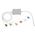
PhysioFlow® PF07 Q-Link™
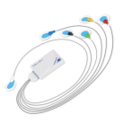
PhysioFlow® Enduro™ Features
PhysioFlow® Q-Link™ Features
.Small Size: 115 x 85 x 18 mm
.Light Weight: Less than 200g (with batteries)
.6 thoracic surface electrodes
.Advanced adaptative filter for noise cancellation (HD-Z™)
.High performance AA batteries or rechargeable AA batteries, 6 hours autonomy
.24 hours MMC memory, USB or wireless download
.Real time wireless monitoring. Range is 100 meters
.Works with PhysioFlow® PF v2 MS Windows™-based software for display, data analysis, and storage
.Minimum computer configuration:
Windows 7, 8, 10
Processor: 2 ghz
Ram : 2GB
Hard Drive 500 MB Free
15″ screen 1280 x 1024
.Recommended computer configuration:
Windows 10, 11
Processor: 2.3 ghz
Ram : 4GB
Hard Drive 5 GB Free
15″ screen 1280 x 1024
.Small Size: 126 x 96 x 20 mm
.Light Weight: Less than 200g
.6 pre-gelled thoracic surface electrodes
.Advanced adaptative filter for noise cancellation (HD-Z™)
.Connections: Patient cable (1 meter), USB cable (about 3 meters) for data transmission and power supply (5V, 300 mA)
.Works with PhysioFlow® PF V2 MS Windows™-based software for display, data analysis, and storage
.Minimum computer configuration:
Windows 7, 8, 10
Processor: 2 ghz
Ram : 2GB
Hard Drive 500 MB Free
15″ screen 1280 x 1024
.Recommended computer configuration:
Windows 10, 11
Processor: 2.3 ghz
Ram : 4GB
Hard Drive 5 GB Free
15″ screen 1280 x 1024
Products

Enduro™ is a portable, battery powered, noninvasive cardiac output device that was designed primarily to be used while a subject or patient is exercising. Enduro™ is unique in that it is the only device that can reliably and repeatedly provide cardiac output during exercise.
A fundamental characteristic of patients with chronic heart failure is an impaired ability to increase cardiac output appropriately with exercise. Many patients are able to improve cardiac output after interventions; cardiac resynchronization therapy and exercise training are directly correlated with this. Quantifying cardiac output can therefore provide important information about these and other interventions. Recent studies have also demonstrated that this information is valuable in predicting outcomes in patients with cardiovascular disease.
PhysioFlow® PF07 Enduro™
Enduro™ is a portable, battery powered, noninvasive cardiac output device that was designed primarily to be used while a subject or patient is exercising. Enduro™ is unique in that it is the only device that can reliably and repeatedly provide cardiac output during exercise.
A fundamental characteristic of patients with chronic heart failure is an impaired ability to increase cardiac output appropriately with exercise. Many patients are able to improve cardiac output after interventions; cardiac resynchronization therapy and exercise training are directly correlated with this. Quantifying cardiac output can therefore provide important information about these and other interventions. Recent studies have also demonstrated that this information is valuable in predicting outcomes in patients with cardiovascular disease.
PhysioFlow® Enduro™ Features
.Small Size: 115 x 85 x 18 mm
.Light Weight: Less than 200g (with batteries)
.6 thoracic surface electrodes
.Advanced adaptative filter for noise cancellation (HD-Z™)
.High performance AA batteries or rechargeable AA batteries, 6 hours autonomy
.24 hours MMC memory, USB or wireless download
.Real time wireless monitoring. Range is 100 meters
.Works with PhysioFlow® PF v2 MS Windows™-based software for display, data analysis, and storage
.Minimum computer configuration:
Windows 7, 8, 10
Processor: 2 ghz
Ram : 2GB
Hard Drive 500 MB Free
15″ screen 1280 x 1024
.Recommended computer configuration:
Windows 10, 11
Processor: 2.3 ghz
Ram : 4GB
Hard Drive 5 GB Free
15″ screen 1280 x 1024

Enduro™ is a portable, battery powered, noninvasive cardiac output device that was designed primarily to be used while a subject or patient is exercising. Enduro™ is unique in that it is the only device that can reliably and repeatedly provide cardiac output during exercise.
A fundamental characteristic of patients with chronic heart failure is an impaired ability to increase cardiac output appropriately with exercise. Many patients are able to improve cardiac output after interventions; cardiac resynchronization therapy and exercise training are directly correlated with this. Quantifying cardiac output can therefore provide important information about these and other interventions. Recent studies have also demonstrated that this information is valuable in predicting outcomes in patients with cardiovascular disease.
PhysioFlow® PF07 Q-Link™
.Small Size: 126 x 96 x 20 mm
.Light Weight: Less than 200g
.6 pre-gelled thoracic surface electrodes
.Advanced adaptative filter for noise cancellation (HD-Z™)
.Connections: Patient cable (1 meter), USB cable (about 3 meters) for data transmission and power supply (5V, 300 mA)
.Works with PhysioFlow® PF V2 MS Windows™-based software for display, data analysis, and storage
.Minimum computer configuration:
Windows 7, 8, 10
Processor: 2 ghz
Ram : 2GB
Hard Drive 500 MB Free
15″ screen 1280 x 1024
.Recommended computer configuration:
Windows 10, 11
Processor: 2.3 ghz
Ram : 4GB
Hard Drive 5 GB Free
15″ screen 1280 x 1024
PhysioFlow® Q-Link™ Features
Is it truly working?
Read the clinical studies reference guide
Is it truly working?
Read the clinical studies reference guide